Hypertension and Martorell’s Ulcer: Role of AI and Telemedicine
A 64-year-old man’s neglected hypertension and diabetes culminated in a painful Martorell’s ulcer—an uncommon ischemic wound of the lower leg. This expanded case study details the clinical presentation, advanced imaging protocols, deep tissue biopsy techniques, AI-driven wound assessment, cloud-based telemedicine follow-up, and expert recommendations for managing such complex ulcers.
Clinical Presentation and Patient History
The patient, an immigrant from Korea who operates a laundromat, presented at Brigham and Women’s Hospital with an 8 × 5 cm full-thickness ulcer over his left ankle, extending 2 cm deep. The wound margins were black, ashen, and purple, with yellow purulent exudate and inflamed red granulation tissue. His vital signs revealed:
- Blood pressure: 215/100 mm Hg (with a three-year history of systolic 160–230 mm Hg, diastolic 95–120 mm Hg).
- HbA1c: 11 % (normal: 4.2–5.6 %), indicating poorly controlled Type 2 diabetes.
- Peripheral pulses: palpable dorsalis pedis and posterior tibial pulses bilaterally.
- Sensory exam: intact light touch, proprioception, and monofilament testing—ruling out diabetic neuropathy.
The patient admitted non-adherence to prescribed regimens: metformin for diabetes, hydrochlorothiazide and losartan for hypertension, and a statin for hyperlipidemia. He stood >8 hours/day at work, but had no history of deep vein thrombosis or significant trauma.
Diagnostic Imaging and Laboratory Workup
Initial bloodwork revealed leukocytosis (white blood cell count 16,000/µL) and elevated C-reactive protein (68 mg/L). Plain radiographs of the ankle demonstrated soft-tissue swelling without periosteal reaction or cortical erosions. Suspecting osteomyelitis, clinicians ordered a 3 Tesla MRI with the following protocol:
- T1-weighted spin-echo (repetition time/echo time: 600/12 ms)
- T2-weighted fat-saturated axial and sagittal sequences
- Contrast-enhanced imaging after intravenous gadobutrol (0.1 mmol/kg)
The MRI identified a soft-tissue defect with no marrow edema or cortical breach, effectively ruling out osteomyelitis. Contrast images confirmed patent tibial arteries and no deep vein thrombosis, reducing concern for large-vessel occlusion.
Deep Wedge Biopsy and Histopathology
A 4 mm punch biopsy of the wound edge was initially non-diagnostic. The team proceeded with a deep wedge biopsy (1.5 cm depth, 2 cm width), sampling epidermis, dermis, subcutaneous fat, and small arteriolar branches. Histological analysis showed:
- Marked arteriolosclerosis: concentric smooth muscle hyperplasia and hyaline deposition in wall of small arteries (media thickness increased by 50 %).
- Fibrinoid necrosis of the tunica intima.
- Extensive inflammatory infiltrate with neutrophils and macrophages.
- Areas of ischemic necrosis in subcutaneous fat lobules.
These findings, combined with the clinical scenario, confirmed the diagnosis of a Martorell’s ulcer—an ischemic ulcer caused by chronic uncontrolled hypertension leading to arteriolar narrowing.
Microbiological Cultures and Secondary Infection
Deep-tissue cultures grew Serratia marcescens and Enterococcus faecalis, both opportunistic Gram-negative and Gram-positive pathogens. Sensitivity testing recommended treatment with:
- Ceftazidime (2 g IV every 8 h) targeting Serratia.
- Ampicillin (2 g IV every 4 h) for Enterococcus.
The antibiotic regimen was de-escalated from broad-spectrum coverage after culture results, reducing risk of antimicrobial resistance.
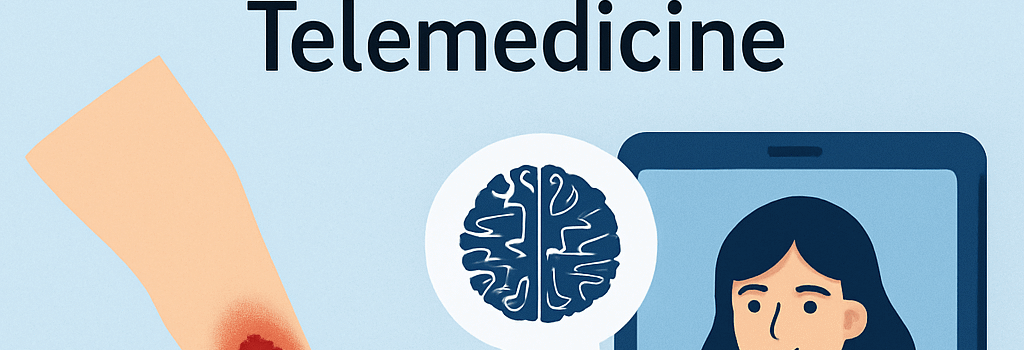
AI-Driven Wound Assessment and Predictive Modeling
In collaboration with the hospital’s digital medicine unit, serial wound photographs were processed through a convolutional neural network (CNN) trained on over 10,000 annotated ulcer images. The AI model provided:
- Automated wound segmentation with 92 % Dice coefficient accuracy.
- Volumetric measurement estimates (± 5 % error).
- Healing trajectory prediction, flagging a 20 % risk of poor closure without surgical intervention.
“AI-enabled imaging allowed the care team to quantify wound area changes objectively and optimize timing for debridement and grafting.” — Dr. Anita Gupta, Vascular Surgeon, Brigham and Women’s Hospital
Cloud-Based Telemedicine and Remote Monitoring
Post-discharge, the patient was enrolled in a cloud-based wound care platform. A HIPAA-compliant mobile app enabled:
- Daily photo uploads of the graft site using a smartphone camera (12 MP sensor).
- Automated alerts when AI detected ≥ 5 % increase in area or signs of exudate.
- Teleconferencing visits with a wound care nurse specialist via WebRTC protocol.
This digital approach improved adherence and enabled early detection of graft failure, demonstrating the promise of telehealth in chronic wound management.
Surgical Management and Skin Grafting
The patient underwent three staged debridement procedures under spinal anesthesia (bupivacaine 0.5 %), removing necrotic tissue with a powered dermabrader (oscillation 1,500 rpm). A split-thickness skin graft (0.012 inch thickness) was harvested from the contralateral thigh using an air-powered dermatome. Negative pressure wound therapy (−125 mm Hg) was applied post-grafting for 5 days to optimize graft adherence.
Optimizing Medical Management and Adherence
Following recovery, the patient was re-educated on pharmacotherapy and enrolled in a digital pill dispenser program with Bluetooth connectivity and adherence reminders. His revised medication plan included:
- Losartan/HCTZ fixed-dose combination (100/25 mg once daily).
- Metoprolol succinate extended-release (50 mg once daily).
- Metformin XR (1,000 mg twice daily).
- Rosuvastatin (20 mg at bedtime).
Home blood pressure monitoring (automated oscillometric device with Bluetooth sensor) and continuous glucose monitoring (CGM) were linked to the telemedicine platform, enabling real-time data review by the care team.
Expert Opinions and Guidelines
“Martorell’s ulcer remains under-recognized. Early biopsy and strict blood pressure control are paramount to prevent progression to full-thickness ischemic wounds.” — Dr. Samuel Lee, Professor of Dermatology, Harvard Medical School
According to the latest American Heart Association guidelines (2025), achieving target blood pressure < 130/80 mm Hg and HbA1c < 7 % reduces risk of microvascular complications, including ischemic ulcers.
Implications for Future Practice
This case underscores the synergy between traditional clinical methods and digital health innovations:
- Deep tissue biopsy: gold standard for diagnosing small-vessel ischemic ulcers.
- AI imaging: enhances objectivity in wound assessment and healing predictions.
- Cloud telemedicine: fosters adherence and early intervention.
As technology evolves, integrating machine learning, advanced imaging, and remote monitoring will be crucial in managing complex chronic wounds effectively.
Additional Resources
- NEJM Clinical Problem-Solving: Martorell’s Ulcer Case Study, Jul 2025.
- AHA/ACC Hypertension Guidelines 2025.
- FDA Guidance on Digital Health Technologies for Remote Patient Monitoring.