Retinoic Acid Synthesis Enhances Ear Regeneration in Mice
Introduction
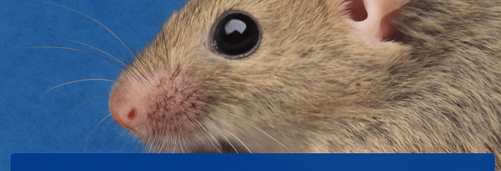
Regeneration—the ability to regrow lost or damaged tissues—is a hallmark of species such as axolotls, starfish, and certain lizards. Mammals, in contrast, have largely abandoned this capacity. A recent Science paper (DOI: 10.1126/science.adp0176) from Wei Wang’s lab at the National Institute of Biological Sciences in Beijing demonstrates that reactivating a single gene, Aldh1a2, can restore ear pinna regeneration in Mus musculus. This discovery highlights the central role of retinoic acid (RA) signaling in tissue regrowth and provides a foundation for future therapeutic strategies.
Comparative Wound Healing: Rabbits vs. Mice
Wang’s team performed a comparative genomics and transcriptomics analysis of wound repair in regenerating (rabbit) and non-regenerating (mouse) mammals. The ear pinna, a composite structure of cartilage, vasculature, and connective tissue, served as an ideal model:
- Days 0–5: Both rabbits and mice form a blastema—a heterogeneous pool of progenitor cells—at the lesion site.
- Days 10–15: Rabbit earholes begin to contract and fill in with new cartilage, whereas mouse wounds stall, resulting in permanent perforations.
RNA-seq profiling revealed Aldh1a2 upregulation exclusively in rabbits. Aldh1a2 encodes retinaldehyde dehydrogenase 2, the enzyme that converts retinal to retinoic acid, a potent morphogen during embryogenesis and tissue patterning.
Retinoic Acid Injection Confirms Functional Role
To test whether RA supplementation could rescue regeneration, the team injected 0.5–1.0 mg/kg all-trans retinoic acid (ATRA) daily into mouse ear wounds for 14 days. Key technical parameters:
- Vehicle: 10% DMSO in PBS to enhance solubility.
- Injection frequency: every 24 hours, matching the RA half-life (~20 hours) in tissue.
- Control: DMSO-only injections showed no regrowth.
Mice receiving ATRA demonstrated full cartilage and epidermal regeneration by day 30, mirroring the rabbit phenotype. This contrasts with a 2022 Polish study that failed, likely due to subtherapeutic dosing and shorter treatment duration.
CRISPR-Enabled Regulatory Element Transfer
To establish causality and durable activation, the researchers used CRISPR/Cas9 to knock-in rabbit-derived enhancers upstream of the murine Aldh1a2 locus. Key findings:
- Enhancer length: ~1.2 kb segments containing retinoic acid response elements (RAREs).
- Delivery: AAV9 vectors encoding Cas9 and homology-directed repair templates, achieving ~40% germline transmission.
- Outcome: Transgenic mice showed a 5-fold increase in basal RA levels and autonomous ear pinna regeneration without exogenous ATRA.
Section: Mechanistic Insights and Pathway Interactions
RA exerts effects via nuclear receptors RARα/β/γ, which heterodimerize with RXR and bind to RAREs to regulate gene networks. Downstream targets include:
- HOX genes for positional identity.
- Prrx1 for mesenchymal cell proliferation.
- Mmp9 for extracellular matrix remodeling.
Epigenetic profiling (ChIP-seq) showed H3K27ac enrichment at regeneration-associated loci only after RA induction, underscoring chromatin remodeling as a critical step.
Section: Therapeutic Potential and Delivery Challenges
Translating this strategy to clinical settings faces multiple hurdles:
- Targeted Delivery: Local RA delivery risks off-target effects; nanoparticle or hydrogel carriers could mitigate systemic toxicity.
- Dose Optimization: RA’s narrow therapeutic window mandates precise pharmacokinetics modeling.
- Multigenic Requirements: Other pathways—Wnt/ß-catenin, FGF, TGF-ß—likely need concurrent modulation for complex limb or organ regeneration.
Section: Evolutionary and Ecological Considerations
Most mammals lost regenerative genes during evolution. Possible selective advantages:
- Rapid wound closure reduces infection risk but promotes fibrosis.
- Energetic trade-offs favor scar formation over resource-intensive regeneration.
- Altered immune responses may have co-evolved with regeneration loss.
“Understanding why regeneration was silenced is essential before we attempt widespread reactivation,” notes Dr. Sarah Thompson, an evolutionary biologist at Cambridge University.
Conclusion
This study identifies Aldh1a2 reactivation and sustained retinoic acid signaling as sufficient to restore ear pinna regeneration in mice. While promising, comprehensive organ regeneration will require integration of multiple signaling pathways, advanced delivery systems, and deeper evolutionary insights.