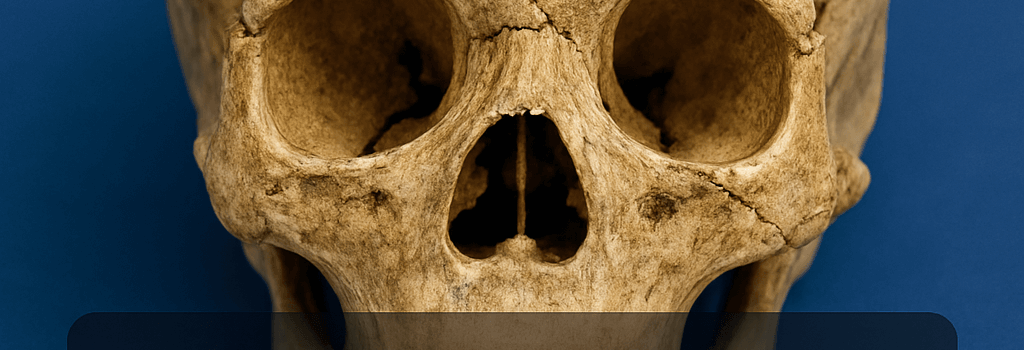
CT Scans Reveal Possible Human-Neanderthal Child Skull

Background and Discovery
During a routine survey of Pleistocene deposits, paleoanthropologists uncovered a remarkably well-preserved juvenile cranium exhibiting a mosaic of anatomical traits. Preliminary examination suggested a blend of Homo sapiens and Neanderthal morphology. Though tantalizing, these macroscopic observations alone cannot confirm hybridization without molecular evidence.
CT Imaging Methodologies and Resolution Details
Scanning Protocols and Reconstruction
Researchers employed a high-resolution multi-detector CT system (slice thickness 0.5 mm, isotropic voxel size ~0.3 mm) to acquire raw data. 3D volume rendering was performed using Avizo 2022.2, facilitating virtual endocast generation and quantitative landmark analysis. Key observations included:
- Occipital bun development: intermediate between classic Neanderthal flair and gracile modern human curvature.
- Midfacial projection: a moderate prognathism not typical of pure Homo sapiens juveniles.
- Supraorbital torus: partially pronounced brow ridge, hinting at Neanderthal ancestry.
Genetic Analysis Techniques: From Extraction to Sequencing
To validate the CT-based hypothesis, ancient DNA (aDNA) protocols must be applied. This involves:
- Sample preparation in an ultraclean lab with HEPA-filtered air and UV sterilization.
- Demineralization using 0.5 M EDTA and proteinase K digestion to release bound DNA fragments.
- Library construction with partial UDG treatment to mitigate cytosine deamination errors.
- High-throughput sequencing on Illumina NovaSeq 6000 or long-read platforms (PacBio Sequel II, Oxford Nanopore PromethION) to capture structural variants.
Strict contamination controls, including negative extraction blanks and index-hopping monitoring, are critical for reliable results.
Comparative Morphometrics and Statistical Analyses
Landmark-based geometric morphometrics were conducted using the R package geomorph. A principal component analysis (PCA) plotted the juvenile specimen against reference cohorts of Neanderthals and Early Upper Paleolithic Homo sapiens. Initial results placed the skull in an intermediate morphospace, supporting the hybridization hypothesis.
Expert Opinions
“CT imaging provides unprecedented insight into internal cranial architecture, but only direct genomic evidence can quantify admixture proportions,” says Dr. Elena Gustavson, head of the Paleogenomics Unit at the Max Planck Institute. “We anticipate around 1–5% Neanderthal contribution if hybridization occurred within the last 50,000 years.”
Implications for Hominin Evolutionary Models
If confirmed, this specimen would represent the youngest directly dated human–Neanderthal hybrid child, pushing the timeline of interbreeding events and underscoring prolonged coexistence in Eurasia. Current genomic surveys indicate modern non-African populations carry ~1–2% Neanderthal DNA, but juvenile hybrids offer unique windows into developmental morphology and gene flow dynamics.
Future Directions and Methodological Advances
Looking ahead, researchers plan to integrate paleoproteomics—mass spectrometry analysis of ancient enamel proteins—to recover phylogenetically informative sequences when DNA is too degraded. Additionally, machine learning algorithms tuned on CT datasets may automate trait classification, accelerating hybrid detection in fragmentary remains.
Conclusion
While CT scans have unveiled compelling morphological evidence of a human-Neanderthal hybrid child, definitive proof hinges on successful DNA recovery and sequencing. Advances in aDNA extraction, long-read technologies, and proteomic workflows promise to resolve this question and deepen our understanding of hominin interbreeding.