First-Time Capture of Plant Cell Wall Construction: A Dynamic Process Uncovered
Introduction: A Breakthrough in Plant Biology
For decades, researchers have understood the static endpoints of plant cell wall formation, but the dynamic process of its assembly remained elusive. Thanks to a novel imaging platform, scientists have now captured the very moment when a plant cell wall is built. This groundbreaking observation opens the door not only to a deeper understanding of plant structure but also to innovative agricultural practices and biotechnological applications.
Overcoming Imaging Challenges: From Fragile Protoplasts to Low Phototoxicity
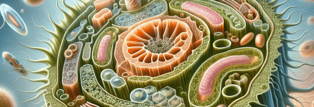
Plant cells normally sport a robust cell wall built primarily of cellulose microfibrils interwoven with complex polysaccharides like hemicellulose and pectin. However, in the absence of this protective layer, cells—referred to as protoplasts—become extremely fragile. Their vulnerability, combined with light-sensitivity, has traditionally thwarted long-term microscopic observations. Researchers faced several technical hurdles:
- Fragility of Protoplasts: Maintaining cell viability over several hours under a microscope was a significant challenge.
- Phototoxicity: Standard microscopy uses high-intensity light, which damages these delicate cells.
- Fluorescence Limitations: Cellulose naturally lacks fluorescence, making direct visualization impossible without appropriate markers.
To address these issues, the research team, led by experts at Rutgers University, designed a custom imaging setup that minimized damage while providing the temporal resolution necessary to capture the wall-building process over a 24-hour period.
Technical Innovations: Custom-Built Imaging Platform and TIRFM
Central to this achievement is the use of Total Internal Reflection Fluorescence Microscopy (TIRFM). Unlike traditional microscopy techniques, TIRFM selectively illuminates a thin section of the sample, dramatically reducing the risk of phototoxicity and allowing longer observation periods. Key technical specifications include:
- Programmable LED Light Source: Automatically deactivates between imaging cycles to further protect the cells from light damage.
- Temperature Control: A system maintaining a consistent 18° C (64° F), which is crucial for optimal cell function during prolonged observation.
- Specialized Fluorescent Markers: Instead of conventional tags that are either toxic or insufficiently specific, the team linked peptide segments from cellulose-binding enzymes to nontoxic green fluorescent markers, enabling high-fidelity imaging of cellulose deposition.
This innovative setup not only captures the process in real time but also lays the groundwork for future enhancements, such as three-dimensional imaging and multimodal fluorescent tagging of enzymes involved in cellulose biosynthesis.
Ordering Chaos: Revealing the Two-Step Process of Cell Wall Assembly
Prior to this study, models of cell wall construction imagined a continuous extrusion of long cellulose fibers. However, the observed process was far more intricate. Initially, the cell produces very short strands of cellulose that appear to drift randomly across the cell surface. Upon encountering one another, these strands merge into longer fibers—comparable to tentacle-like projections—that subsequently intertwine to form an organized matrix. This two-step assembly process prompts new questions about the underlying mechanics:
- Is the merging of cellulose fibers driven by directed, energy-dependent mechanisms, or is it purely stochastic, occurring through random collisions?
- What regulatory proteins and enzymes orchestrate the precise timing and spatial arrangement of these fibers?
Insights from experts like Rutgers’ Eric Lam and Shishir Chundawat emphasize that this unexpected complexity challenges classical textbook diagrams and paves the way for a new understanding of cell wall biosynthesis.
Biotechnological Implications: Enhancing Biomass Yield and Crop Robustness
The implications of visualizing the real-time construction of cell walls are significant. By identifying and manipulating the key genes and enzymes that regulate this process, scientists envision the possibility of engineering plants with higher biomass yield. Such modifications could:
- Boost the efficiency of agricultural land use, increasing the output per hectare.
- Enhance the structural robustness of crops, potentially reducing susceptibility to environmental stress and pests.
- Lower the production costs for biofuels and biochemicals, thus driving down overall expenses in sustainable energy production.
Much like deconstructing and refining the components of a car engine to improve performance, the systematic disruption and subsequent analysis of each gene in the cellulose biosynthesis pathway will provide detailed insights into optimizing plant growth and productivity.
Expert Opinions: Insights from the Front Lines of Research
According to Professor Eric Lam from Rutgers University, “We knew the starting and finishing points of cell wall formation, but witnessing the intermediate steps completely upended our understanding. It’s a reminder that nature often surprises us when we develop the right tools to observe it.” Similarly, biologist Shishir Chundawat stresses, “This discovery not only reshapes our theoretical models but opens new avenues for practical applications in agriculture and renewable energy.” Their expert assessments underscore the transformative potential of this research in bridging basic science with applied technology.
Future Directions: 3D Imaging and Beyond
While this breakthrough represents a significant milestone, many questions remain. The current time-lapse only captured the final deposition of cellulose at the cell surface. Future research will focus on creating three-dimensional visualizations of the entire biosynthetic pathway. By tagging other critical enzymes with distinct fluorescent markers, researchers hope to map out the intricate choreography of biochemical interactions that lead to cell wall formation.
Furthermore, integrating machine learning algorithms to analyze vast amounts of imaging data could reveal subtle patterns and regulatory feedback loops that would be invisible to the human eye. This fusion of advanced imaging and data analytics represents the next frontier in plant science, with potential ripple effects across biotechnology, synthetic biology, and agricultural engineering.