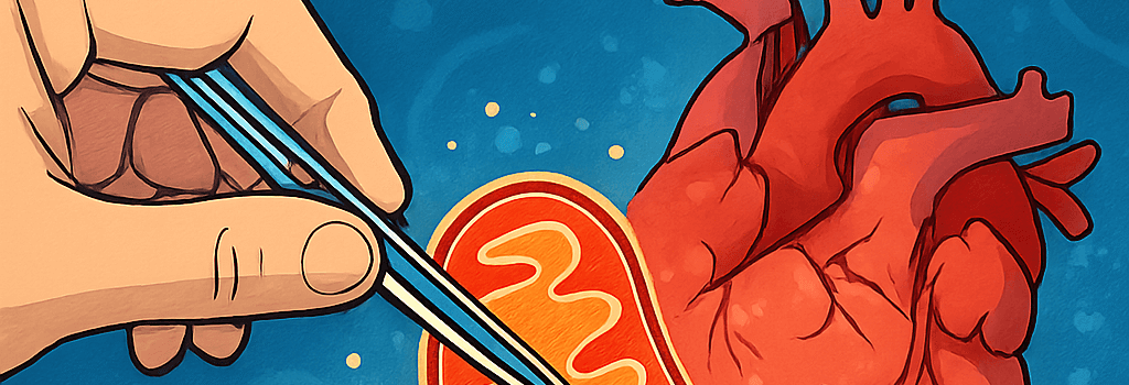
Engineering Mitochondria for Organ Repair

Researchers worldwide are refining mitochondrial transplantation techniques to rescue ischemia-damaged organs, from hearts and brains to kidneys and livers. Advances in isolation protocols, delivery systems, and biobanking could soon enable off-the-shelf cellular power units for regenerative medicine.
Historical Breakthroughs in Mitochondrial Transplantation
Nearly two decades ago, James McCully at Boston Children’s Hospital pioneered the first live demonstration of mitochondrial rescue in a pig heart. Using differential centrifugation—700 × g for 10 min to remove nuclei and cellular debris, followed by 10,000 × g for 15 min to pellet mitochondria—his team isolated organelles with >85% membrane potential (assessed via JC-1 fluorescence). A single injection of ~1 × 10⁸ functional mitochondria in Krebs buffer restored contractility in an ischemic pig heart within minutes, reducing infarct volume by 40% relative to controls.
Mechanisms of Mitochondrial Uptake and Integration
At the cellular level, transplanted mitochondria employ actin-mediated endocytosis and macropinocytosis to enter target cells. Once inside, they evade mitophagy by modulating PINK1/Parkin signaling and rapidly fuse with the endogenous network through OPA1/MFN1-2-dependent pathways. Recent super-resolution microscopy studies (STED) show donor mitochondria integrating into the host’s reticulum within 30–60 min, boosting ATP synthesis rates by 25–50% (measured via Seahorse XF assays).
Clinical Applications: From Cardiac Surgery to Neurological Repair
- Pediatric Cardiac Support
- Ischemic Stroke Intervention
- Organ Preservation for Transplantation
Pediatric Cardiac Support
Between 2015 and 2018, McCully and cardiac surgeon Sitaram Emani treated 10 infants undergoing complex congenital heart surgery. Using a filtered mitochondrial extract (2 mg tissue yielding ~2 × 10⁸ organelles per mL), they injected directly into the myocardium post–cardiopulmonary bypass. Eight infants weaned off extracorporeal life support within 48 h (vs. historical control average of nine days). A 2025 follow-up study expanded the cohort to 25 patients, confirming safety and demonstrating a 35% reduction in postoperative inflammatory cytokines (IL-6, TNF-α).
Ischemic Stroke Trials
Melanie Walker’s team at UW Medicine conducted a Phase I safety trial (2024) in four patients with acute middle cerebral artery occlusion. After thrombectomy, they delivered 5 mL of mitochondrial suspension (~5 × 10⁷ organelles/mL) via microcatheter distal to the lesion. No procedure-related adverse events were reported. A Phase II trial launching in late 2025 will quantify mitochondrial homing using ^13C-labeled tracers and MR spectroscopic imaging to confirm functional integration.
Organ Preservation and Donation Optimization
Giuseppe Orlando’s Wake Forest group evaluated mitochondria-enhanced hypothermic machine perfusion of pig kidneys (n=4) vs. controls (n=3). They infused 1 × 10⁹ organelles in oxygenated University of Wisconsin solution at 4 °C. Post-perfusion histology showed a 60% decrease in TUNEL-positive cells, while ex vivo respirometry indicated a twofold increase in state-3 respiration. A 2025 preprint extends these findings to human livers, demonstrating improved bile production and reduced transaminase release.
Challenges in Mitochondria Isolation and Storage
Scaling mitochondria production for clinical use demands robust GMP-compliant protocols. Differential centrifugation yields high-purity organelles but is labor-intensive. Cryopreservation with 5% DMSO results in a 50% drop in membrane potential; optimized lyophilization techniques are under investigation. Koning Shen (UC Berkeley) warns that maintaining RCR (respiratory control ratio) above 0.7 post-thaw remains a key hurdle for off-the-shelf mitochondrial banks.
Emerging Delivery Techniques
Innovative approaches are in development to target mitochondria precisely and protect them during transit. Lipid nanoparticle carriers functionalized with targeting peptides (e.g., mitochondrial targeting sequence MTS conjugates) show enhanced uptake efficiency. Ultrasound-mediated microbubble cavitation is being tested to transiently permeabilize vascular endothelium and improve organelle delivery in vivo. AI-driven peptide design is accelerating the discovery of novel mitochondria-penetrating sequences with high specificity and low immunogenicity.
Future Perspectives and Regulatory Pathways
Before FDA approval, mitochondrial therapies must clear IND requirements, including sterility (<0.25 EU/mL), endotoxin limits, and validated potency assays. Biobanking initiatives aim to cryostore diverse mitochondrial subtypes—cardiac, neuronal, renal—under a unified quality management system. Venture-backed startups are exploring automated isolation platforms powered by microfluidic sorting. Navdeep Chandel (Northwestern) and Lance Becker (Northwell Health) emphasize the need for mechanistic clarity, proposing multiomics studies to map mitochondrial-nuclear cross-talk post-transplant.
“We’re so much at the beginning—we don’t know how it works,” Becker says. “But we know it’s doing something mighty interesting.”
Conclusion
Mitochondrial transplantation is poised to revolutionize organ repair and preservation. As researchers refine isolation, delivery, and storage, the vision of universal mitochondria banks and off-the-shelf rejuvenation therapies moves closer to reality. Continued interdisciplinary collaboration will be essential to translate these cellular powerhouses into standard clinical tools.